Introduction
The human scalp, a complex and vital component of our integumentary system, is subject to a variety of medical conditions that can significantly impact an individual’s health, aesthetic appearance, and psychological well-being. Among these, alopecia and tinea capitis stand out as two prevalent yet fundamentally different conditions that often cause concern due to their external manifestations—hair loss and scalp abnormalities. Although their symptoms may overlap superficially, especially in early stages, their origins, clinical presentations, diagnostic approaches, and treatment strategies diverge sharply.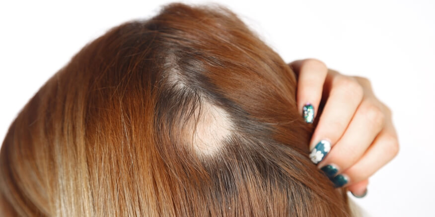
This comprehensive discussion, curated by the esteemed Free Source Library platform (freesourcelibrary.com), aims to delineate the distinctions and commonalities between alopecia and tinea capitis in a nuanced manner, emphasizing scientific accuracy, clinical relevance, and practical implications for healthcare professionals, students, and affected individuals. Recognizing these differences is quintessential for accurate diagnosis, tailored treatment protocols, and effective management of these conditions to restore not only scalp health but also improve quality of life.
Furthermore, an in-depth exploration of the neuroimmunological, genetic, environmental, microbiological, and therapeutic dimensions of these disorders will be provided, supported by current scientific literature, to facilitate a thorough understanding of their pathophysiology, epidemiology, and the latest advancements in medical and alternative interventions.
Understanding Alopecia
Alopecia, broadly defined as hair loss, encompasses a wide spectrum of disorders characterized by partial or complete baldness. The complexity of alopecia lies in its multifactorial etiology, involving genetic predispositions, autoimmune processes, hormonal fluctuations, and environmental factors. The diversity of its subtypes reflects distinct pathogenic mechanisms, clinical presentations, and tailored approaches to management.
Types of Alopecia
Alopecia Areata: Autoimmune-mediated Patchy Hair Loss
Alopecia areata is classified as an autoimmune disorder wherein the immune system erroneously targets hair follicle structures, leading to localized patches of non-scarring hair loss. It tends to manifest suddenly, often without warning, and can affect individuals of any age, although it is most prevalent in young adults. The characteristic presentation involves one or multiple round, smooth, bald patches, which may appear suddenly and can vary in size.
The pathogenesis involves a complex interplay between genetic susceptibility and environmental stimuli, such as stress or viral infections, which precipitate an immune response against hair follicle antigens. Histologically, a perifollicular lymphocytic infiltrate surrounded by healthy hair follicles characterizes this condition. The hair follicles in affected areas are often in the anagen phase prematurely, resulting in hair shedding. Some cases are transient and resolve spontaneously, whereas others progress.
Androgenetic Alopecia: Patterned Hair Thinning
Androgenetic alopecia, colloquially known as male-pattern baldness or female-pattern hair loss, is the most common form of chronic hair thinning. It results from a complex interaction between genetic predisposition and hormonal influences, notably androgens like dihydrotestosterone (DHT). It is characterized by a patterned hair loss with distinctive features—receding hairlines and balding crowns in men, and diffuse thinning over the vertex in women.
Genetically, genes involved span multiple chromosomes, and their expression influences hair follicle sensitivity to hormones. The role of androgens involves binding to androgen receptors within hair follicles, leading to miniaturization—a gradual transformation from terminal to vellus-like hair. This process results in a thinning appearance over time, with hair shaft diameters decreasing, and a reduction in hair density.
Alopecia Totalis and Universalis: Total Loss of Hair
Alopecia totalis is an advanced form of alopecia areata characterized by complete scalp hair loss, while alopecia universalis extends this process to body hair, including eyebrows and eyelashes. Both conditions are autoimmune in origin, with immune-mediated destruction of hair follicles across the affected areas. These forms often present with significant psychological distress and require comprehensive management strategies.
Diagnosis of Alopecia
Diagnosing alopecia involves a meticulous clinical examination and supportive laboratory investigations. The clinician assesses the pattern, extent, and characteristics of hair loss, checking for signs of scarring, inflammation, or systemic disease indicators. Dermoscopy, or scalp microscopy, can reveal specific features such as exclamation mark hairs in alopecia areata or miniaturized follicles in androgenetic alopecia.
In certain cases, scalp biopsy remains the gold standard, allowing microscopic evaluation of follicular architecture, inflammatory infiltrates, and fibrosis. Blood tests might be performed to exclude systemic conditions like thyroid abnormalities or iron deficiency anemia, which can exacerbate hair loss.
Management Strategies for Alopecia
Treatment options are tailored according to the type and severity of alopecia. For autoimmune disorders such as alopecia areata, corticosteroids—topical, intralesional, or systemic—are often employed to suppress immune responses. Immunotherapy with contact allergens or JAK inhibitors shows promise, although these are used experimentally or in refractory cases.
Androgenetic alopecia management focuses on slowing progression and promoting hair regrowth. Minoxidil, a topical vasodilator, is approved for both sexes, increasing blood flow to hair follicles and stimulating growth. Finasteride, a 5-alpha-reductase inhibitor, reduces androgen conversion, particularly effective in men. Hair transplantation remains an option for suitable candidates, involving grafting healthy follicles from donor areas.
Emerging therapies, such as platelet-rich plasma (PRP) injections and low-level laser therapy, are gaining popularity, although their long-term efficacy requires further validation through clinical trials.
Unveiling Tinea Capitis
Tinea capitis, colloquially known as scalp ringworm, is a superficial fungal infection predominantly caused by dermatophytes—fungi that digest keratinized tissue. Unlike alopecia, which is primarily telogen or miniaturization of hair follicles due to internal factors, tinea capitis involves an infectious process that directly damages hair shafts and scalp tissue.
Though it can affect all ages, it is most common in children between the ages of 3 and 10 years. Its contagious nature and environmental persistence, especially in crowded settings such as schools, foster rapid spread if hygiene measures are not strictly maintained.
Etiology and Pathogenesis
The primary causative agents include dermatophyte genera such as Trichophyton and Microsporum. These fungi colonize keratinized tissues, producing enzymes like keratinases that facilitate invasion of hair shafts and scalp epithelium. The infection triggers an inflammatory response characterized by follicular pustules, scaling, and hair breakage.
The mode of transmission typically involves direct contact with infected humans or animals, as well as indirect contact with fomites such as brushes, hats, bedding, or towels. Importantly, environmental contamination is common, with fungal spores capable of surviving on surfaces for extended periods.
Clinical Manifestations
Early Symptoms
The initial phase often presents as scalp pruritus, mild erythema, and patchy hair loss. The affected areas might be covered with fine scaling that may be mistaken for dandruff. Over time, signs evolve, and distinct patterns emerge.
Characteristic Signs
- Scaly patches: These areas of the scalp show circumscribed, erythematous, scaly plaques with varying degrees of inflammation.
- Hair breakage: Hair shafts in infected zones become brittle, leading to broken hairs that appear as ‘black dots’ at the scalp surface.
- Inflammatory lesions: Some cases, especially in more aggressive infections, display pustules, erythema, and swelling.
- Kerion: Severe inflammatory nodular lesions, often with suppuration, are termed kerions, sometimes mistaken for abscesses but representing a hypersensitive immune response.
- Transmission signs: Presence of infected pets or close contact individuals suggests recent or ongoing exposure.
Diagnostic Techniques
Clinical assessment provides initial clues, but microbiological confirmation is essential. Fungal cultures from scalp scrapings or plucked hairs identify the specific dermatophyte involved. Microscopy using potassium hydroxide (KOH) preparations reveals fungal hyphae and spores within the sample.
A Wood’s lamp examination can assist by causing fluorescing of certain Microsporum species, aiding rapid presumptive diagnosis. PCR-based molecular diagnostics are increasingly used for precise species identification, which can influence treatment choices.
Treatment and Preventive Measures
Pharmacologic Interventions
Systemic antifungal agents are the mainstay, with griseofulvin, terbinafine, and itraconazole being the most common. The choice of drug depends on factors such as age, severity, dermatophyte species, and drug tolerability.
A typical course lasts between 6 to 8 weeks, ensuring eradication of the infectious agent. It’s essential to adhere strictly to the prescribed duration to prevent recurrence and resistance development.
Adjunct Hygiene and Care
- Regular shampooing with medicated antifungal shampoos such as selenium sulfide or ketoconazole helps reduce fungal burden and transmission.
- Cleaning bedding, hats, brushes, and clothing thoroughly to eliminate spores.
- Avoiding sharing personal items and maintaining good scalp hygiene.
- Managing close contacts and treating infected pets if applicable.
Comparison Table of Key Differences Between Alopecia and Tinea Capitis
| Feature | Alopecia | Tinea Capitis |
|---|---|---|
| Cause | Genetic, autoimmune, hormonal factors | Fungal dermatophyte infection |
| Appearance | Smooth, bald patches, diffuse thinning | Scaly, inflamed patches with hair breakage |
| Contagious? | No | Yes, highly contagious |
| Transmission | Not contagious | Contact with infected individuals or animals |
| Diagnosis | Clinical examination, biopsy, blood tests | Microscopy, fungal culture, Wood’s lamp |
| Treatment | Topical/systemic immune modulators, minoxidil, medications | Oral antifungals, hygiene measures |
| Prognosis | Variable; some recover spontaneously, others need ongoing treatment | Often curable with proper therapy, but prone to recurrence |
Emerging Perspectives and Future Directions
Advances in molecular biology, immunology, and pharmacology continue to shape the understanding and management of alopecia and tinea capitis. Emerging therapies, such as JAK inhibitors for alopecia areata, offer promise in reversing autoimmune hair loss. On the other hand, novel antifungal agents with broader spectrum and fewer side effects are under development for dermatophyte infections, including resistant strains.
Research into the microbiome of the scalp reveals complex interactions between host, fungi, bacteria, and other microbes, potentially influencing the onset and progression of both conditions. Personalized medicine approaches, considering individual genetic makeup and microbial profiles, could revolutionize treatment efficacy and prevention strategies.
Furthermore, public health initiatives emphasizing hygiene education, pet management, and early intervention will be crucial for controlling the spread of tinea capitis, especially in vulnerable populations like children.
Conclusion
Understanding the nuanced differences between alopecia and tinea capitis is fundamental for clinicians and patients alike. While both conditions can produce alarming hair loss and scalp disruptions, their root causes, clinical signs, and management pathways are distinct. Accurate diagnosis is paramount, achieved through a combination of clinical evaluation and laboratory investigations. Implementation of tailored treatments ensures the best outcomes, minimizes complications, and enhances quality of life.
As ongoing research unravels the complex biology of these scalp conditions, continuous adaptation of diagnostic tools and therapies promises improved prognosis. The importance of informed awareness, precise diagnostics, and effective intervention cannot be overstated—particularly in safeguarding scalp health within community settings.
For those seeking further information or authoritative sources, references such as the National Institutes of Health and peer-reviewed journals provide extensive research articles supporting these insights.