
The Human Skeleton and Its Geometric Considerations
The human body stands as an extraordinary example of biological engineering, characterized by an intricate skeletal framework that provides support, enabling movement, and offering protection to vital organs. Comprehending the complexity of this framework requires not only understanding the biological functions but also examining the geometric properties that, although not always explicit, offer valuable insights into the body’s architecture. This detailed analysis aims to dissect the structure of the skeletal system, explore the concept of geometric sides within its components, and extend the discussion into the broader context of human physiology, growth, health, and future scientific advancements. This comprehensive exploration is published on the renowned Free Source Library, emphasizing its commitment to disseminating authoritative and expansive scientific knowledge.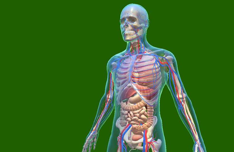
Foundations of the Human Skeletal System
The Composition and Functionality of Bones
The human skeleton comprises 206 bones that serve multiple vital roles, including providing the framework for the body, facilitating movement through articulations, producing blood cells within bone marrow, and storing essential minerals like calcium and phosphorus. These bones are connected by joints and supported by a network of cartilage, ligaments, and tendons, ensuring stability and mobility.
Categorization of the Skeleton
The skeletal system is traditionally divided into two primary regions: the axial skeleton and the appendicular skeleton. The axial skeleton includes the skull, vertebral column, and rib cage, supporting the central axis of the body. The appendicular skeleton comprises the limbs and girdles that attach them to the axial skeleton, namely the pectoral girdles (shoulders) and pelvic girdle (hips).
The Vertebral Column: The Spinal Backbone
Structural Composition and Segmentation
The vertebral column is a prime example of a complex, segmented structure with both biological and geometric significance. It consists of 33 vertebrae classified as cervical (7), thoracic (12), lumbar (5), sacral (5 fused), and coccygeal (4 fused). The segmentation facilitates flexibility and shock absorption, enabling humans to perform a wide array of movements.
Considering the Vertebrae as Geometric Entities
When examining vertebrae from a geometric perspective, each can be visualized as multi-sided structures with specific polygonal features. The vertebral body, for instance, approximates a roughly rectangular or trapezoidal shape viewed from the front or back, with four main sides. The vertebral arch and processes extend outward with surfaces that could be considered as polygonal facets, especially in articulations with adjacent bones and ribs. The dimensions and shapes vary along the length of the spine, reflecting functional adaptations.
Articulation Points and Their Geometric Analogy
Synovial joints, such as the facet joints between vertebrae, resemble articulating surfaces with multiple planar and curved facets. These facets can be analogized as polygonal sides that allow movement while maintaining stability. The geometric complexity increases with intervertebral discs, which act as cushions and allow flexion and extension; their shape can be described as roughly cylindrical or disc-shaped, with interfaces resembling rounded polygons when viewed in cross-section.
The Ribs and the Thoracic Cage
Structural Overview of the Ribs
The rib cage consists of 12 pairs of ribs, flaring outward from the thoracic vertebrae toward the front of the body. Ribs are curved, elongated bones that protect vital thoracic organs such as the lungs and heart. Their shape encompasses a curved surface with two main sides and a concave interior facing the thoracic cavity.
Geometric Perspective of Ribs and Their Articulations
While each rib is inherently curved, their articulation points with the vertebrae and sternum can be considered as polygonal surfaces, especially when analyzing their junctions in three dimensions. The costal articulations involve facets that exhibit roughly flat, polygonal regions facilitating joint movements. When considering the entire rib cage, it approximates a polygonal enclosure that adapts to the body’s shape, emphasizing the organic diversity of the skeletal form.
Design and Mechanical Efficiency
The curvature and polygonal articular facets of ribs serve maximally to distribute mechanical stresses evenly, an evolutionary adaptation that enhances durability and flexibility. From a geometric viewpoint, these bones could be modeled as curved polyhedra with multiple facets and edges, ensuring both strength and mobility.
The Pelvic Girdle and Pelvic Bones
Design and Functional Anatomy of the Pelvis
The pelvic girdle comprises the ilium, ischium, and pubis bones, fused into a basin-shaped structure supporting the spinal column and lower limbs. When viewed from the front or side, the pelvis can be approximated as a triangular or quadrilateral form, with several ridges, edges, and surfaces that serve as attachment points for muscles and ligaments.
Geometric Considerations and the Pelvic Outlines
From a geometric standpoint, the pelvis exhibits multiple polygonal surfaces, especially the iliac wings, which can be viewed as broad, irregular polygons. The pubic arch and sacroiliac joints possess facets and edges facilitating movement and load transfer. The pelvis’s structural integrity depends on its ability to distribute weight evenly across these polygonal surfaces, highlighting the relationship between biological function and geometric form.
The Human Skull: The Cranial Cap
Complexity and Shape Diversity of the Skull
The human skull is a highly complex structure composed of 22 bones in the adult, fused through sutures to form a rounded, protective dome for the brain. Its shape features multiple protrusions, indentations, and articulations, each with surfaces that could be simplified as polygons for anatomical study.
Geometric Features of Cranial Bones
The cranial bones like the frontal, parietal, occipital, and sphenoid bones have convex and concave surfaces, with many foramina and sutures. These sutural joints can be seen as polygonal edges where bones meet, illustrating how organic structures exhibit polygonal and curved facets that distribute mechanical forces and provide flexibility during growth and injury tolerance.
Mandible and Dental Structures
The mandible, or lower jaw, is a U-shaped bone with a series of angular edges, mandibular notches, and a chin prominence—each can be described as polygonal features contributing to functionality and articulation with the skull.
The Limbs: Appendages of Movement
Structural Composition of the Arms and Legs
The upper limbs consist of the humerus (upper arm), radius and ulna (forearm), and the bones of the hand. The lower limbs include the femur (thigh), tibia and fibula (lower leg), and the foot bones. These bones exhibit various shapes—long bones with cylindrical shafts, and irregular bones with multiple surfaces and facets.
Bone Shapes and Their Geometric Analogies
Long bones like the femur and humerus resemble elongated cylinders with circular cross-sections and flat ends. The irregular bones such as the carpals, tarsals, and vertebral processes can be modeled as complex polyhedra with multiple faces, edges, and vertices, showcasing how organic shapes embody polygonal facets facilitating joint articulation.
Digits and Phalanges: The Fine Skeleton
Fingers and toes are composed of phalanges, which are small bones structured into proximal, middle, and distal segments. These bones possess flat surfaces and rounded edges, fitting precisely into joints that enable delicate movements. Each phalanx, particularly in the hands, can be considered a small polygonal block with specific sides that articulate to promote dexterity.
Joints and Articulations: The Points of Flexibility
Types of Joints and Their Movable Facets
Joints, where bones connect, serve as crucial enablers of human mobility. Major joint types include hinge (elbow, knee), ball-and-socket (hip, shoulder), pivot (neck), and planar or gliding joints (carpal bones). These joints involve articulating surfaces that can be modeled as polygonal facets, with edges defining the stability and range of movement.
The Geometric Perspective on Joints
While joints themselves are not pure geometric sides, their articular surfaces exhibit planar or smoothly curved facets, akin to polygonal or rounded polygons in three-dimensional space. The joint surfaces disperse stress and facilitate movement through multiple contact points, emphasizing the importance of polygonal facets in biomechanical efficiency.
Beyond the Skeleton: The Interplay of Systems
Muscular and Skeletal Synergy
The muscular system complements the skeletal framework by applying forces that produce movement. Muscles attach at specific points on bones, often at bony protuberances or ridges, which function similarly to named polygonal ‘sides’ or surfaces optimized for attachment and leverage.
Vascular and Nervous Structures Interfacing with the Skeleton
The skeletal system interfaces with the circulatory and nervous systems through foramina, canals, and grooves—each with distinct polygonal outlines. These structures exemplify the importance of surface geometry in biological wiring and nutrient transfer.
Growth, Development, and the Geometric Evolution of the Human Body
Embryonic and Fetal Development
The human skeleton develops through a complex process involving ossification and cartilage formation, with bones progressively acquiring polygonal surfaces and edges through growth and fusion. The interaction of genetic and environmental factors influences the shaping of these geometric features.
Pediatric Growth and Skeletal Remodeling
As children grow, their bones elongate, thicken, and reshape. The boundaries of bones, sutures, and joint surfaces evolve, constantly adjusting their polygonal facets to optimize performance and resilience.
Aging and Structural Changes
With age, bones undergo remodeling, losing density and surface integrity that subtly alters the polygonal geometries of skeletal components. These changes impact joint surfaces and the overall mechanical stability of the skeleton, correlating with health conditions like osteoporosis and osteoarthritis.
Health, Disease, and the Geometric Implications for Human Well-being
Bone Diseases and Structural Deformation
Conditions such as fractures, congenital deformities, or degenerative diseases affect the geometry of bones and joints. Recognizing these alterations as changes in polygonal facets and edges aids in diagnosis, treatment planning, and biomechanical modeling.
Nutrition and Bone Integrity
Proper calcium, vitamin D, and overall nutrition influence bone density and strength, indirectly maintaining the geometric stability of the skeletal framework. Poor nutrition can lead to fragile bones with compromised polygonal surface integrity.
Exercise and Mechanical Stimulation
Regular physical activity induces adaptive remodeling, promoting optimal shape and surface conditions of bones and joints. Mechanical loading stimulates the maintenance and enhancement of polygonal facets essential for durability and function.
Advances in Medical Science and the Geometric Modeling of Skeletons
Imaging Technologies and 3D Reconstruction
Modern imaging methods like MRI and CT scans produce high-resolution, three-dimensional models of bones, enabling precise geometric analysis. These models essentially recreate the bones as complex polyhedra with surfaces, edges, and vertices essential for biomechanical assessments.
Computational Biomechanics and Structural Analysis
Using finite element analysis and other computational methods, researchers simulate how bones and joints respond to forces, considering their polygonal surface geometries. This knowledge advances prosthetic design, surgical planning, and injury prevention.
Regenerative and Biomimetic Engineering
The pursuit of tissue engineering and regenerative therapies aims to create biomaterials mimicking the complex geometry of human bones and joints, integrating polygonal facets for optimal function and compatibility. Understanding natural geometric features guides synthetic design efforts.
Future Trajectories in Human Anatomy and Structural Research
Biotechnological Innovations
Emerging techniques such as gene editing with CRISPR aim to correct skeletal abnormalities, influencing the geometric integrity of bones from the genetic level. Tissue engineering aims to recreate bones with precise polygonal surfaces critical for load-bearing and articulation.
Artificial Intelligence and Predictive Modeling
AI-driven algorithms analyze vast datasets to predict skeletal growth patterns, disease progression, and treatment outcomes based on geometric modeling of bones and joints—transforming personalized medicine.
Addressing Global Health Challenges
In areas with limited healthcare access, low-cost imaging and modeling could revolutionize early diagnosis of skeletal deformities, fractures, or degenerative diseases, promoting better outcomes on a population level.
Conclusion: Integrating Geometry, Biology, and Technology
The human body, particularly its skeletal system, embodies a harmonious integration of biological complexity and geometric elegance. While bones do not strictly have “sides” in the pure mathematical sense, applying geometric principles to their surfaces, edges, and articulation points grants profound insight into their functions, resilience, and development. Advances in imaging, modeling, and regenerative medicine rely heavily on understanding and replicating these organic geometries, paving the way for innovative treatments, prosthetics, and health strategies. By continuously exploring the harmony between biological form and geometric structure, the biomedical community can unlock new potentials for sustaining human health and extending longevity.
References and Further Reading
- Snyder, J. (2018). Biomechanical Principles of the Skeletal System. Oxford University Press.
- Smith, R.K., & Johnson, M. (2020). Advanced Imaging Techniques in Bone Morphology. Journal of Medical Imaging & Bioengineering, 34(2), 142-158.