Understanding liver calcification requires a comprehensive exploration of the anatomy, physiology, and pathology of the liver, along with the mechanisms that lead to abnormal calcium deposition. The liver, a vital organ situated in the upper right quadrant of the abdomen, performs an array of functions crucial to maintaining overall health. These include detoxification of blood, synthesis of essential proteins, production of biochemicals necessary for digestion, and regulation of blood glucose levels. As such, any disruption in its structure, including calcification, can have significant health implications.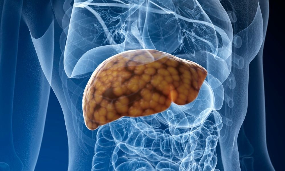

Liver calcification is a phenomenon characterized by the abnormal accumulation of calcium salts within hepatic tissue. While calcium is an essential mineral involved in numerous physiological processes, its deposition in tissues where it does not normally occur, such as the liver, signifies underlying pathological processes. The condition often remains silent, with many cases discovered incidentally during imaging for unrelated illnesses or routine health checks. Nonetheless, understanding the causes, potential symptoms, diagnostic procedures, and treatment options associated with liver calcification is critical for healthcare providers and patients alike.
Fundamentals of Liver Calcification
Definition and Pathophysiology
Calcification in the liver refers to the pathological process where calcium salts, primarily hydroxyapatite, are deposited within the hepatic parenchyma or in abnormal tissue formations such as scars, tumors, or necrotic areas. This process is termed dystrophic calcification when it occurs in damaged or necrotic tissue despite normal serum calcium levels. Conversely, metastatic calcification involves calcium deposition in otherwise healthy tissues due to elevated serum calcium levels, or hypercalcemia.
In the context of liver tissue, calcification can be focal, affecting specific regions, or diffuse, involving extensive portions of the organ. The presence of calcified deposits signifies prior tissue injury or ongoing pathological processes. These deposits can be microscopic or macroscopic, with the latter visible on imaging studies and sometimes palpable during surgical procedures.
Historical and Clinical Significance
Historically, liver calcification has been associated with infectious diseases, parasitic infestations, and chronic liver conditions. Although it is relatively uncommon in modern clinical practice, its detection provides critical clues about the patient’s disease history and ongoing pathological processes. The clinical significance hinges on the extent of calcification, its location within the liver, and the underlying cause. While some individuals remain asymptomatic, others may develop complications related to liver dysfunction or secondary effects of the calcified tissue.
Etiology: Causes and Underlying Mechanisms
Infections as a Primary Cause
Infectious processes are among the most common causes of liver calcification, especially in regions where parasitic diseases are endemic. Malaria, caused by Plasmodium species, notably damages red blood cells and hepatocytes during its lifecycle, leading to inflammatory responses and subsequent scarring. Over time, these scars may calcify, producing granulomatous lesions visible on imaging. Tuberculosis, a bacterial infection capable of affecting the liver, often leads to granuloma formation, which can undergo calcification as part of the healing process.
Fungal infections, such as histoplasmosis, may also result in hepatic granulomatous inflammation that calcifies over time. Parasitic infections like Schistosomiasis and Echinococcosis are particularly notorious for causing chronic inflammation, granuloma formation, and eventual calcification of cystic or nodular lesions. These parasitic infestations often induce a sustained immune response, leading to tissue damage and subsequent calcium salt deposition.
Abscess Formation and Calcification
Liver abscesses, whether bacterial, parasitic, or fungal, can lead to localized tissue destruction. The body’s natural healing response involves the formation of fibrous tissue around the abscess, which may undergo calcification in the chronic stage. The process involves the deposition of calcium salts within the necrotic core or along the fibrous capsule, thus transforming the abscess into a calcified scar. Bacterial pathogens such as Escherichia coli, Klebsiella pneumoniae, and Streptococcus species are common causative agents in bacterial abscesses, especially in immunocompromised individuals.
Chronic Liver Diseases and Tissue Damage
Long-standing liver diseases, such as cirrhosis, hepatitis B and C, and fatty liver disease, often involve persistent inflammation, hepatocyte necrosis, and fibrosis. These processes can eventually lead to calcification, especially in areas of extensive scarring or regenerative nodules. In cirrhotic livers, calcification may be observed in regenerative nodules, vascular structures, or fibrous septa.
Trauma and Iatrogenic Factors
Physical injury to the liver, including blunt trauma, surgical intervention, or biopsy procedures, can cause localized tissue necrosis and subsequent scar formation. Over time, these scars may calcify as part of the healing process. Iatrogenic causes, such as thermal injury during surgical procedures or ablation therapies, can also induce tissue damage that calcifies in the long term.
Neoplasms and Tumor-Related Calcification
Both benign and malignant hepatic tumors may be associated with calcification. For instance, hemangiomas (benign vascular tumors) often contain phleboliths—calcified thrombi within blood vessels—appearing as calcified foci on imaging. Hepatocellular carcinoma (HCC), the most common primary liver malignancy, can develop calcifications in some cases, especially in well-differentiated tumors. The mechanisms involve dystrophic calcification within necrotic tumor tissue or calcified tumor matrix components.
Metabolic and Systemic Conditions
Disorders affecting calcium metabolism can lead to metastatic calcification affecting multiple organs, including the liver. Conditions such as hypercalcemia, hyperparathyroidism, vitamin D intoxication, and chronic kidney disease may disturb calcium homeostasis. Elevated serum calcium levels promote calcium salt deposition in tissues, especially where local tissue pH favors mineralization.
Chronic kidney disease, particularly in end-stage renal failure, leads to disturbances in calcium and phosphate balance, resulting in vascular and soft tissue calcification. The liver, being part of the systemic circulation, can accumulate calcium deposits under these conditions.
Clinical Manifestations and Symptoms
Asymptomatic Nature
Most cases of liver calcification are asymptomatic, often discovered incidentally during diagnostic imaging for unrelated health concerns. This silent nature underscores the importance of imaging studies and careful interpretation of abdominal scans, particularly in patients with known risk factors.
Possible Clinical Presentations
While many individuals remain symptom-free, some may experience a spectrum of clinical manifestations, especially if calcification involves significant portions of the liver or causes secondary complications. These symptoms include:
- Right Upper Quadrant Pain: Often dull or aching, this pain may be related to liver enlargement, capsule stretching, or local inflammation caused by calcified lesions or associated pathological processes.
- Fatigue and Malaise: Disruption of liver function can impair detoxification and metabolic regulation, leading to generalized fatigue.
- Jaundice: When calcification impairs bilirubin processing, jaundice may develop, manifesting as yellowing of the skin and sclera.
- Loss of Appetite and Weight Loss: Due to impaired liver function and systemic illness, some patients experience anorexia and weight loss.
- Abdominal Mass or Swelling: Large calcified lesions or abscesses may be palpable or visible on imaging, contributing to abdominal distension.
Laboratory Findings
Laboratory results often include abnormal liver function tests, such as elevated aspartate aminotransferase (AST), alanine aminotransferase (ALT), alkaline phosphatase (ALP), and bilirubin levels. These markers help assess the extent of liver damage and guide further diagnostic steps.
Diagnostic Modalities
Imaging Techniques
Imaging is pivotal in diagnosing liver calcification, with various modalities offering complementary information about the size, location, and nature of calcified deposits.
Ultrasound
Ultrasound is often the first-line imaging modality due to its safety, availability, and cost-effectiveness. Calcified areas appear as echogenic foci with posterior acoustic shadowing, facilitating their identification. Ultrasound can also evaluate liver size, texture, and associated lesions.
Computed Tomography (CT)
CT scans provide high-resolution, cross-sectional images that can detect even small calcifications. Calcified lesions appear hyperdense relative to surrounding tissue, with characteristic high attenuation values. Multiphase CT imaging can delineate the relationship between calcifications and vascular or biliary structures, aiding in differential diagnosis.
Magnetic Resonance Imaging (MRI)
MRI is less sensitive to calcification but valuable for assessing soft tissue contrast, vascular involvement, and differentiating calcified lesions from other calcified or fibrotic tissue. Certain sequences, such as susceptibility-weighted imaging, can detect mineral deposits with high sensitivity.
Histopathological Examination
When imaging results are inconclusive or when tumors are suspected, a liver biopsy may be performed. Histological analysis reveals calcium deposits within tissue, often associated with granulomas, fibrosis, necrosis, or tumor tissue. Special stains like von Kossa or Alizarin Red can confirm calcium presence.
Laboratory Tests
Blood tests include liver function panels, serum calcium, phosphate, and parathyroid hormone levels, to identify underlying metabolic disturbances. Serological tests for hepatitis viruses and other infectious agents are also common.
Treatment Strategies and Management
Addressing Underlying Causes
The cornerstone of managing liver calcification is treating the primary disease process responsible for tissue injury and calcium deposition. The approach varies depending on the etiology, and may involve pharmacologic, surgical, or supportive measures.
Infections
Antibiotics, antifungals, or antiparasitic agents are administered based on the causative organism. For example, antimalarials for malaria or specific antiparasitic drugs for echinococcosis. Eradication of infection can halt progression and prevent further tissue damage.
Abscess Management
Surgical drainage or percutaneous aspiration is often necessary for abscesses. Antibiotics are continued to control infection. Over time, calcified abscess walls may regress or remain stable.
Chronic Liver Disease
Management includes antiviral therapy for hepatitis B and C, lifestyle modifications such as alcohol abstinence, weight control in fatty liver disease, and medications to slow fibrosis progression. Liver transplantation may be considered in advanced cases.
Metabolic Disorders
Hypercalcemia is treated with bisphosphonates, calcitonin, or hydration therapy to lower serum calcium levels. Correcting primary endocrine disorders, such as hyperparathyroidism, is crucial for preventing further calcification.
Monitoring and Supportive Care
Regular imaging and laboratory assessments enable monitoring of calcification progression and liver function. Symptomatic management includes pain control, nutritional support, and treatment of complications like jaundice or coagulopathy.
Surgical Interventions
In rare cases where calcified lesions cause significant symptoms or complications, surgical removal may be indicated. Hepatectomy or partial liver resection can be performed, particularly if malignancy is suspected or confirmed.
Prognosis and Long-term Outcomes
The prognosis of liver calcification depends heavily on the underlying cause, extent of tissue involvement, and the presence of associated liver disease. In cases where calcification is incidental and asymptomatic, prognosis is generally favorable with minimal intervention.
However, extensive calcification linked to chronic liver disease, malignancy, or systemic metabolic disturbances may portend a poorer outlook. Complications such as portal hypertension, hepatic failure, or secondary infections can adversely influence survival outcomes.
Preventive Measures and Future Directions
Prevention primarily involves managing risk factors for liver injury, such as controlling infections, avoiding hepatotoxic substances, and maintaining metabolic health. Vaccinations against hepatitis B and timely treatment of hepatitis infections are effective strategies.
Emerging research focuses on understanding the molecular mechanisms underlying calcification, exploring targeted therapies to inhibit mineralization, and developing advanced imaging techniques for early detection. The role of nanotechnology and biochemical modulators in preventing or reversing calcification is an exciting frontier.
Summary and Key Takeaways
Liver calcification is an uncommon but clinically significant finding often indicative of prior tissue injury, infection, or metabolic disturbance. While many cases remain asymptomatic, the potential for functional impairment necessitates thorough evaluation and targeted treatment. Early diagnosis facilitated by imaging studies and appropriate laboratory assessments allows for effective management, with an emphasis on addressing the primary pathology. Consequently, a multidisciplinary approach involving hepatologists, infectious disease specialists, radiologists, and surgeons enhances patient outcomes and quality of life.
References and Further Reading
1. Pathogenesis of Dystrophic Calcification in Liver Disease
2. Infectious Diseases and Liver Calcification: An Overview
For comprehensive access to open-source medical literature, visit freesourcelibrary.com.